Nanosurgery with femtosecond lasers

Photodisruption in turbid tissue with 100-fs and 200-ps laser pulses, at APS Centennial Meeting 1999 (Atlanta, GA), Friday, March 26, 1999:
We compare the potential of 100-fs and 200-ps laser pulses for photodisruptive surgery on the surface and in the bulk of turbid tissue. Water, human epidermis cultures, and pig skin were used as tissue models. In our technique, tightly-focused femtosecond and picosecond laser pulses are nonlinearly absorbed, vaporizing tissue in the focal volume. We find that there are several advantages in using femtosecond pulses for photodisruption. The breakdown threshold is lower and the energy deposition is more deterministic for 100-fs pulses compared to 200-ps pulses. In human skin culture we observe... Read more about Photodisruption in turbid tissue with 100-fs and 200-ps laser pulses
Single neuron dissection in C. elegans by femtosecond laser pulses, at Photonics West 2006 (San Jose, CA), Saturday, January 21, 2006:
Using tightly-focused, 2-5 nJ femtosecond laser pulses we disrupt cellular material in the nematode worm C. elegans. Due to the nonlinear absorption of laser light, the dissection has submicrometer resolution, yielding surgery within the bulk of the worm without incision. The low energy of the pulses minimizes collateral damage. This technique permits subcellular surgery on live animals and opens a wide range of neurobiological questions to study in vivo.

We severed individual dendrites of a thermosensory neuron without damaging nearby neurons. Quantification of the resulting behavioral...

Read more about Single neuron dissection in C. elegans by femtosecond laser pulses
Subcellular surgery and nanosurgery, at Physics Colloquium, University of Kentucky (Lexington, KY), Thursday, October 25, 2007:
We use femtosecond laser pulses to manipulate sub-cellular structures inside live and fixed cells. Using only a few nanojoules of laser pulse energy, we are able to selectively disrupt individual mitochondria in live bovine capillary epithelial cells, and cleave single actin fibers in the cell cytoskeleton network of fixed human fibro-blast cells. We have also used the technique to micromanipulate the neural network of C. Elegans, a small nematode. Our laser scalpel can snip individual axons without causing any damage to surrounding tissue, allowing us to study the function of individual... Read more about Subcellular surgery and nanosurgery
Manipulating cells using ultrashort laser pulses, at Wednesday Night Research Seminar, Harvard University (Cambridge, MA), Wednesday, September 9, 2009:
We use femtosecond laser pulses to manipulate sub-cellular structures inside live and fixed cells. Using only a few nanojoules of laser pulse energy, we are able to selectively disrupt individual mitochondria in live bovine capillary epithelial cells, and cleave single actin fibers in the cell cytoskeleton network of fixed human fibro-blast cells. We have also used the technique to micromanipulate the neural network of C. Elegans, a small nematode. Our laser scalpel can snip individual axons without causing any damage to surrounding tissue, allowing us to study the function of individual... Read more about Manipulating cells using ultrashort laser pulses
Subcellular surgery and nanosurgery, at Western Washington University (Bellingham, WA), Tuesday, May 13, 2014:
We use femtosecond laser pulses to manipulate sub-cellular structures inside live and fixed cells. Using only a few nanojoules of laser pulse energy, we are able to selectively disrupt individual mitochondria in live bovine capillary epithelial cells, and cleave single actin fibers in the cell cytoskeleton network of fixed human fibro-blast cells. We have also used the technique to micromanipulate the neural network of C. Elegans, a small nematode. Our laser scalpel can snip individual axons without causing any damage to surrounding tissue, allowing us to study the function of individual... Read more about Subcellular surgery and nanosurgery
Nanosurgery in live cells using ultrashort laser pulses, at 2005 SPIE Photonics West Conference (San Jose, CA), Tuesday, January 25, 2005:
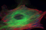
We selectively disrupted the cytoskeletal network of a live bovine capillary endothelial cell using ultrashort laser pulses. We image the microtubules in the cytoskeleton of the cultured cells using green fluorescent protein. The cells are placed on a custom-built inverted fluorescence microscope setup, using a 1.4 NA oil-immersion objective to both image the cell and focus the laser radiation into the cell samples. The laser delivers 100-fs laser pulses centered at 800 nm at a repetition rate of 1 kHz; the typical energy delivered at the sample is 1–5nJ. The fluorescent image of the cell is... Read more about Nanosurgery in live cells using ultrashort laser pulses
Probing cell mechanics with femtosecond laser pulses, at Photonics West 2007 (San Jose, CA), Sunday, January 21, 2007:
We use femtosecond laser pulses to selectively disrupt the cytoskeleton of a living cell and probe its mechanical properties. Our nanosurgery setup is based on a home-built fluorescence microscope with an integrated femtosecond laser. We severed single actin bundles inside live cells to probe the local dynamics of the cytoskeleton and correlate it to global changes in cell shape. Simultaneous cutting and imaging allows us to study immediate cellular response with several hundred-nanometer spatial and less than 500-ms time resolution. The targeted actin bundle retracts rapidly after laser... Read more about Probing cell mechanics with femtosecond laser pulses
Subcellular surgery and nanosurgery, at Special Laser Seminar / NCCR MUST Seminar, ETH Zürich (Zürich, Switzerland), Thursday, February 23, 2012:
We use femtosecond laser pulses to manipulate sub-cellular structures inside live and fixed cells. Using only a few nanojoules of laser pulse energy, we are able to selectively disrupt individual mitochondria in live bovine capillary epithelial cells, and cleave single actin fibers in the cell cytoskeleton network of fixed human fibro-blast cells. We have also used the technique to micromanipulate the neural network of C. Elegans, a small nematode. Our laser scalpel can snip individual axons without causing any damage to surrounding tissue, allowing us to study the function of individual... Read more about Subcellular surgery and nanosurgery
Photodisruption in biological samples using femtosecond laser pulses, at Photonic West Conference (San Jose, CA), Tuesday, January 23, 2001:
Tightly focused femtosecond laser pulses can be absorbed nonlinearly inside biological materials, vaporizing tissue in the focal volume. High intensity is achieved at the focus with relatively low energy, minimizing collateral damage and unwanted heat deposition. We focus 100-fs laser pulses using a high numerical aperture microscope objective in the bulk of the skin tissue, disrupting micrometer-sized regions up to 100 mm beneath the sample surface. When the laser beam is focused even deeper into the sample, we observe the formation of filaments in the skin bulk rather than spherical sub-... Read more about Photodisruption in biological samples using femtosecond laser pulses
Subcellular surgery and nanosurgery, at CIMIT/Lester Wolfe Workshop on Femtosecond Microscopy & Microsurgery, Wellman Center for Photomedicine (Boston, MA), Tuesday, April 18, 2006:
We use femtosecond laser pulses to manipulate sub-cellular structures inside live and fixed cells. Using only a few nanojoules of laser pulse energy, we are able to selectively disrupt individual mitochondria in live bovine capillary epithelial cells, and cleave single actin fibers in the cell cytoskeleton network of fixed human fibro-blast cells. We have also used the technique to micromanipulate the neural network of C. Elegans, a small nematode. Our laser scalpel can snip individual axons without causing any damage to surrounding tissue, allowing us to study the function of individual... Read more about Subcellular surgery and nanosurgery
Subcellular surgery and nanosurgery, at Phi Beta Kappa Lecture, Pomona College (Claremont, CA), Tuesday, April 15, 2008:
We use femtosecond laser pulses to manipulate sub-cellular structures inside live and fixed cells. Using only a few nanojoules of laser pulse energy, we are able to selectively disrupt individual mitochondria in live bovine capillary epithelial cells, and cleave single actin fibers in the cell cytoskeleton network of fixed human fibro-blast cells. We have also used the technique to micromanipulate the neural network of C. Elegans, a small nematode. Our laser scalpel can snip individual axons without causing any damage to surrounding tissue, allowing us to study the function of individual... Read more about Subcellular surgery and nanosurgery
Subcellular surgery and nanosurgery, at PR-LSAMP Role Model Series, UPR Rio Piedras (Rio Piedras, PR), Friday, February 19, 2010:
We use femtosecond laser pulses to manipulate sub-cellular structures inside live and fixed cells. Using only a few nanojoules of laser pulse energy, we are able to selectively disrupt individual mitochondria in live bovine capillary epithelial cells, and cleave single actin fibers in the cell cytoskeleton network of fixed human fibro-blast cells. We have also used the technique to micromanipulate the neural network of C. Elegans, a small nematode. Our laser scalpel can snip individual axons without causing any damage to surrounding tissue, allowing us to study the function of individual... Read more about Subcellular surgery and nanosurgery
High Throughput Poration of Mammalian Cells using Femtosecond Laser-activated Plasmonic Substrates, at Tokyo Metropolitan University (Tokyo, Japan), Thursday, January 29, 2015:
We present a new cell transfection method that uses femtosecond laser-excited localized surface plasmons (LSPs) on a nanostructured micropyramid array. Our gold-layered micropyramids have nano-apertures at the apex to form high local electric field enhancements, or “hot spots.” These hot spots form microbubbles that temporarily perforate mammalian cell (HeLa S3) membranes and allow dye molecules and plasmid vectors to diffuse through the membrane openings. We optimize our laser parameters for successful poration and high cell viability. Our nontoxic, efficient, and scalable technique... Read more about High Throughput Poration of Mammalian Cells using Femtosecond Laser-activated Plasmonic Substrates
Femtosecond laser dissection of neurons in C. elegans, at Industrial Outreach Program, Harvard University (Cambridge, MA), Wednesday, April 27, 2005:
Tightly-focused femtosecond laser pulses of a few nanojoules sever individual dendrites in the nematode worm C. elegans. Quantification of the resulting behavioral deficits identifies the contribution of the dissected structures. The dissection has submicrometer resolution with no collateral damage, permitting precise studies on live animals. Future work include an examination of the molecular basis of neurodegeneration that has application to diseases such as Parkinsons and Alzheimers.

Pages